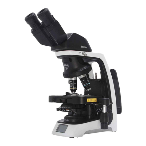
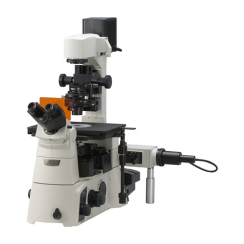
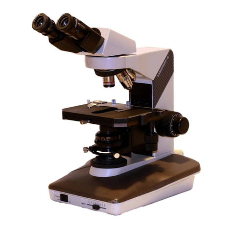
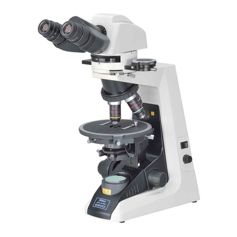
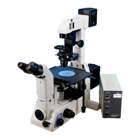
Nikon Eclipse Ci-E User manual

Upright Motorized Microscope
Instruction Manual
M569E 17.9.NF.9 (1/3)
*M569EN09*


Introduction
i
Introduction
Thank you for purchasing a Nikon product.
This instruction manual is written for users of the Nikon ECLIPSE Ci-E microscope. To ensure correct usage, read this
manual carefully before operating this product.
●No part of this manual may be reproduced or transmitted in any form without prior written permission from Nikon.
●The contents of this manual are subject to change without notice.
●The equipment described in this manual may differ from the actual product in its appearance.
●Although every effort has been made to ensure the accuracy of this manual, errors or inconsistencies may remain. If
you note any points that are unclear or incorrect, please contact your nearest Nikon representative.
●Some of the equipment described in this manual may not be included in the set you have purchased.
●If you intend to use any other equipment with this product, read the manual for that equipment too.
●If this equipment is used in a manner not specified by the manufacturer, the protection provided by the equipment may
be impaired.

Introduction
ii
Contents of the Manual
The manual for ECLIPSE Ci-E consists of the following contents. There is also a "Bright-field Microscopy Quick Guide",
provided as a separate document.
This manual: Instructions
Safety Precautions
Microscopy Procedures
Bright-field Microscopy
Phase Contrast Microscopy
Simple Polarizing Microscopy
Sensitive Polarization Microscopy
Epi-fluorescence Microscopy
Individual Operations
Assembly
Troubleshooting
Maintenance and Storage
Specifications and Safety Standards
Bright-field Microscopy Quick Guide
Symbols Used in This Manual
The following symbols are used in this manual.
Symbols for Safety
WARNING
CAUTION
Highlights important information that should be noted for safety. Read “Safety Precautions” for details.
Other Symbols
Indicates information you should note or comply with to prevent defects or malfunction of this product.
Indicates information you should be aware of in using this product, as well as other useful information.

Table of Chapters
iii
Chap. 1-1
Chap. 1-2
Chap. 1-3
Chap. 1-4
Chap. 1-5
Chap. 2
Chap. 3
Chap. 5
Chap. 4
Chap. 6
Table of Chapters (See next page for the detailed contents.)
Introduction
Contents of the Manual
Symbols Used in This Manual
Safety Precautions/
Notes on Handling the Product
Microscopy Procedures
Bright-field Microscopy
Microscopy Procedures
Phase Contrast Microscopy
Microscopy Procedures
Simple Polarizing Microscopy
Microscopy Procedures
Sensitive Polarization Microscopy
Microscopy Procedures
Epi-fluorescence Microscopy
Individual Operations
Assembly
Troubleshooting
Maintenance and Storage
Specifications and Safety Standards

Contents
iv
Contents
Introduction .........................................................................................................................i
Contents of the Manual ....................................................................................................ii
Symbols Used in This Manual ..........................................................................................ii
Table of Chapters.............................................................................................................. iii
Safety Precautions........................................................................................................... vii
WARNING and CAUTION Symbols ...............................................................................vii
Meaning of Symbols Used on the Product.....................................................................vii
WARNING ............................................................................................................... viii
CAUTION ..................................................................................................................ix
Notes on Handling the Product.......................................................................................... x
Microscopy Procedures.......................................................................... 1
1 Bright-field Microscopy..............................................................................................1
1.1 System Configuration and Controls ................................................................... 1
1.2 Bright-field Microscopy Procedure ..................................................................... 3
2 Phase Contrast Microscopy ....................................................................................11
2.1 System Components and Controls ...................................................................11
2.2 Phase Contrast Microscopy Procedure ........................................................... 13
3 Simple Polarizing Microscopy .................................................................................18
3.1 System Configuration and Controls ................................................................. 18
3.2 Simple Polarizing Microscopy Procedure ........................................................ 20
4 Sensitive Polarization Microscopy...........................................................................25
4.1 System Configuration and Controls ................................................................. 25
4.2 Sensitive Polarization Microscopy Procedure.................................................. 27
5 Epi-fluorescence Microscopy ..................................................................................32
5.1 System Configuration and Controls ................................................................. 32
5.2 Epi-fluorescence Microscopy Procedure ......................................................... 34
Individual Operations............................................................................ 39
1 Adjusting the Brightness of a Diascopic Image.......................................................39
1.1 Adjustment with the Dia-illumination Brightness Control Knob........................ 39
1.2 Adjustment with ND Filters............................................................................... 39
2 Focusing on the Specimen (Vertical Stage Movement)...........................................40
2.1 Focus Knob Rotation and Stage Movement .................................................... 41
2.2 Number of Focus Knob Turns and Distance of Stage Travel........................... 41
2.3 Adjusting the Rotating Torque of the Coarse Focus Knob............................... 42
2.4 Refocusing ....................................................................................................... 42
2.5 Position Exchange of the Fine Focus Knob ..................................................... 43
3 Bringing the Specimen into the Optical Path (Horizontal Stage Movement) ...........44
3.1 Knob Rotation and Stage Movement ............................................................... 44
3.2 Adjusting the Knob Heights.............................................................................. 44
3.3 Adjusting the Knob Rotation Torque................................................................. 44
4 Adjusting the Diopter...............................................................................................45
5 Focusing and Centering the Condenser .................................................................46
6 Adjusting the Aperture Diaphragm ..........................................................................47
6.1 Adjusting the Aperture Diaphragm Using the Condenser Scale...................... 47
6.2 Adjusting the Aperture Diaphragm Using the Centering Telescope................. 47
7 Selecting a Condenser............................................................................................48

Contents
v
8 Adjusting the Field Diaphragm ................................................................................48
9 Switching the Optical Path of the Tube ...................................................................49
9.1 Light Distribution............................................................................................... 49
9.2 Disabling the Clicking of the Optical Path Switching ....................................... 49
10 Adjusting the Binocular Section ..............................................................................50
10.1 Adjusting with the Binocular Section of the Tube............................................. 50
10.2 Using the Eyelevel Riser .................................................................................. 50
11 Using at Lowered Position of Stage (Spacer for Nosepiece) ..................................50
12 Switching Objectives ...............................................................................................51
12.1 Basic Operation................................................................................................ 51
12.2 Switching between Two Objectives (Toggle).................................................... 52
13 Configuring the Motorized Functions ......................................................................54
13.1 Configuring the Motorized Swing-out Condenser ............................................ 54
13.2 Changing the Initial Brightness of Dia-illumination LED .................................. 55
13.3 Reverse Rotation Lock (Rotation from Address 1 to 6) ................................... 56
14 Oil Immersion ..........................................................................................................57
15 Water Immersion .....................................................................................................58
16 Tips for Phase Contrast Microscopy .......................................................................58
17 Tips for Epi-fluorescence Microscopy .....................................................................60
17.1 Switching Excitation Methods .......................................................................... 61
17.2 Selecting Filters................................................................................................ 62
18 Capturing Images....................................................................................................64
18.1 Photomicroscopy.............................................................................................. 64
18.2 Tips on Microscope Settings for Photomicroscopy .......................................... 65
Assembly................................................................................................ 67
1 ECLIPSE Ci-E System Configuration......................................................................67
2 Assembly for Bright-field Microscopy ......................................................................68
3 Assembly for Phase Contrast Microscopy ..............................................................73
4 Assembly for the Simple Polarizing Microscopy .....................................................73
5 Assembly for Sensitive Polarization Microscopy .....................................................74
6 Assembly for Epi-fluorescence Microscopy ............................................................75
7 Attaching a Camera ................................................................................................77
Troubleshooting..................................................................................... 79
1 Optical System and Operation ................................................................................79
1.1 General............................................................................................................. 79
1.2 Epi-fluorescence Microscopy ........................................................................... 83
1.3 Phase Contrast Microscopy ............................................................................. 84
2 Electrical System.....................................................................................................85
2.1 General............................................................................................................. 85
2.2 Epi-fluorescence Microscopy ........................................................................... 86
Maintenance and Storage ..................................................................... 87
1 Cleaning ..................................................................................................................87
1.1 Cleaning Lenses............................................................................................... 87
1.2 Cleaning Parts Other than the Lens................................................................. 87
1.3 Cleaning Immersion Oil .................................................................................... 88

Contents
vi
1.4 Decontaminating the Product........................................................................... 88
2 Storage....................................................................................................................88
3 Regular Inspections (Charged) ...............................................................................88
Specifications and Safety Standards................................................... 89
1 Microscopy (Principles) ...........................................................................................89
2 Performance Properties ..........................................................................................89
3 Physical Properties .................................................................................................90

Safety Precautions
vii
Safety Precautions/Notes on Handling the Product
Safety Precautions
To ensure correct and safe operation, read this manual before using this product.
WARNING and CAUTION Symbols
Although this product is designed and manufactured to be completely safe during use, incorrect usage or failure to follow
the safety instructions provided may cause personal injury or property damage. To ensure correct usage, read this manual
carefully before using this product. Do not discard this manual and keep it handy for easy reference.
Safety instructions in this manual are marked with the following symbols to indicate their importance. For your safety,
always follow the instructions marked with these symbols.
Symbol Description
WARNING Disregarding instructions marked with this symbol may lead to serious injury or death.
CAUTION Disregarding instructions marked with this symbol may lead to injury or property damage.
Meaning of Symbols Used on the Product
When appearing on this product, the symbols below indicate the need for caution at all times during use. Read the
relevant instructions in this manual before attempting to use or adjust any part to which the symbol has been affixed.
Biohazard
This symbol is affixed to the front of the stand of this product, to call your attention to the following:
●WARNING: Using this product may constitute a biohazard risk if a sample comes into contact with this
product.
●To avoid biohazard contamination, do not touch the contaminated portion with bare hands.
●Decontaminate the contaminated part in accordance with the standard procedure specified for your
laboratory.

Safety Precautions
viii
Safety Precautions/Notes on Handling the Product
WARNING
1 Do not disassemble.
Disassembling this product may result in electric
shock or malfunction. Malfunction and damage
due to disassembling or modification are
unwarranted.
Do not disassemble parts other than those
described in this manual. If you experience
problems with this product, contact your nearest
Nikon representative.
2 Read the instruction manuals carefully.
To ensure safety, thoroughly read this manual and
the manuals for other equipment to be used with
this product. Particularly, all warnings and
cautions given at the beginning of each manual
must be observed.
Safety is a top design priority for Nikon products.
Safety is ensured as long as the user observes all
of the warnings and cautions given in the
manuals, and uses the system only for its
intended purpose. However, failure to heed the
warnings and cautions given in the manuals,
subjecting the system to shock or impact, or
attempting to disassemble the system may result
in unexpected accidents and injury.
Product with CI-FL or D-FL Epi-fluorescence
attachment:
The light source used for Epi-fluorescence
microscopy (HG Precentered Fiber Illuminator)
requires special care during handling because of
its characteristics. Be sure to refer to the manual
for the light source being used.
3 Notes on the power cord
Be sure to use the specified power cord. Use of
other power cords may result in malfunction or
fire. This product is classified as having Class I
protection against electric shock. Make sure this
product is connected to an appropriate protective
earth terminal.
Refer to Chapter 6, “2 Performance Properties” fo
r
the specified power cords.
●To prevent electric shock, always turn off the
power switch (press to the “O” position) for the
microscope before connecting or disconnecting
the power cord.
4 Hazards of mercury lamps
(when using a CI-FL or D-FL Epi-fluorescence
attachment)
The light source used with the epi-fluorescence
attachment (HG precentered fiber illuminator)
requires special care during handling because of
its characteristics. For safe and correct use of this
system, carefully read the warnings below. Keep
in mind all potential hazards. Additionally, carefully
read the manual for the illuminator and the manual
from the lamp manufacturer (if provided), then
follow the instructions given therein. Failure to
heed the warnings and cautions given in the
manuals, subjecting the system to shock or
impact, or attempting to disassemble the system
may result in unexpected accidents and injury.
●Ultraviolet light
When lit, mercury lamps radiate ultraviolet light
that can damage the eyes and skin. Direct
viewing of the light may result in blindness.
When changing filter cubes, always turn off the
light source of the Epi-fluorescence
attachment. Leaving the lamp turned on during
filter cube replacement may result in ultraviolet
exposure.
●High-pressure gas
The lamps contain sealed gas under very high
pressure. And the pressure increases when the
lamp is on. If the lamp is scratched, fouled,
subjected to high external pressure or physical
impact, or used beyond its service life, the
sealed gas may leak or the lamp may burst,
resulting in gas inhalation, injury from glass, or
other accidents.
●Heat
When the lamp is lit, the lamp and surroundings
will become extremely hot. Do not touch the
lamp with bare hands or place flammable
materials near the lamp. Failure to comply may
result in burns or fire.
●Designated lamp
Be sure to use the designated lamp. Using
other types of lamps may result in accidents,
including bursting of the lamp.
5 Hazardous sample handling
This product is intended primarily for microscopic
observations and image capture of cells and
tissue set on glass slides.
Check to determine whether a sample is
hazardous before handling. If sample is
hazardous, handle it according to the standard
procedure specified for your laboratory. If the
sample is potentially infectious, wear rubber
gloves and avoid directly touching samples. If
such a sample is spilled onto this product, the
portion must be decontaminated in a safety
manner. Consult your safety supervisor or safety
standard of your facility.
Safety Precautions
ix
Safety Precautions/Notes on Handling the Product
CAUTION
1 Power shutdown
To prevent electric shock and/or malfunction,
always turn off the power switch(es) for this
product and the peripheral devices (press to the
“O” position) and unplug the power cord from the
wall outlet before assembling this product,
connecting or disconnecting cables, or cleaning
this microscope and the objective.
2 Movement of motorized device
With this product, the motorized nosepiece can be
electronically rotated and the condenser top lens
can be electronically swung out. The various
motorized components can also be operated by
the remote controller or from a PC. Various
motorized components can also be operated from
remote controllers and PCs.
To avoid unexpected injuries, note the following in
operating motorized devices.
●Before operation, check the state of the entire
microscope system to ensure safety when
operating the motorized devices.
●Keep your hands and fingers away from the
nosepiece, objectives, stage, and components
and specimen on the stage to avoid injuries
during operation.
3 Prevent contact with water.
Never allow water to come into contact with this
product, and avoid using this product in
circumstances where it may be splashed with
water. Splashing water onto this product may
cause a short circuit, resulting in malfunction or
abnormal heating. If water is splashed onto this
product, immediately turn off the power switches
for this product and the peripheral devices (press
to the “O” position) and remove the power cord
from the receptacle. Then wipe off moisture with a
piece of dry cloth or something similar. If water
enters, stop the use of the product and contact
your nearest Nikon representative.
4 Do not place any object on top of the product.
Do not place any object on top of this product.
5 Cautions on assembling, installing, and
carrying the product
●Take care to avoid pinching your fingers or
hands during product assembly and
installation.
●Scratches or fouling optical components (such
as lens and filters) with fingerprints, etc. will
degrade microscope images. Be careful to
avoid scratches or direct contact with the lens
and filters when assembling.
●The main body weighs approximately 10 kg.
When moving it, hold the main body by the grip
on the back and the recessed portion at the
front bottom side of this product.
●Remove all attachments (if mounted) from the
microscope before moving the microscope.
●Do not place this product in a locker or cabinet.
6 Cautions on use, transportation, and storage
This product must be operated, transported, or
stored in accordance with the following conditions.
Installing this product in a hot, humid location may
result in the formation of mold or condensation on
lenses, impairing performance or causing
malfunctions.
●Operating conditions:
temperature: 0 to +40C, humidity: 60% RH
max. (no condensation)
●Transporting/storage conditions:
temperature: -20 to +60C, humidity: 90% RH
max. (no condensation)
7 Remove any covers from the product before
switching on.
Do not use this product while it is covered with a
piece of cloth, etc., as this will prevent heat
release and result in abnormal heat and create a
fire hazard. Do not cover this product with a piece
of cloth or similar while in use. The system
temperature will rise, resulting in a malfunction.
8 Cautions on sustained observations
To relieve fatigue resulting from long observation
sessions, limit continuous observations to one
hour. Take at least 10 to 15 minutes breaks
between observation sessions. Adjust the layout
of other equipment used and the height of your
chair.
9 Cautions on the disposal of the product
To avoid biohazard risks, dispose of this product
as contaminated equipment in accordance with
the standard procedure specified for your facility.

Notes on Handling the Product
x
Safety Precautions/Notes on Handling the Product
Notes on Handling the Product
1 Handle with care
This product is a precision optical instrument and
requires gentle handling. Avoid subjecting it to
sudden impacts and shocks.
Even relatively minor impacts are capable of affecting
the precision of the objective.
2 Electromagnetic Environment
Before using this product, Nikon recommends
evaluating the electromagnetic environment of the
installation site.
Do not use this product close to strong
electromagnetic radiation sources (example:
unshielded intentional RF sources). They may
interfere with the proper operation of this product.
This product emits weak electromagnetic waves. So
as to avoid degrading the performance of precision
electronic devices, do not install this product near
such devices. If TV or radio reception is affected,
move the TV or radio further from this product.
3 Dirt on the lens
Scratches or fouling optical components (such as
lens and filters) with fingerprints, etc. will degrade
microscope images.
If these parts become dirty, clean them as described
in Chapter 5, “1.1 Cleaning Lenses”.
4 Installation location
This product is a precision instrument. Usage or
storage of this product in an inappropriate
environment may result in malfunction or a
degradation in precision. Consider the following
factors when selecting an installation location:
●Select a location free of vibration. Install this
product on a level surface.
●Install this product at least 10 cm away from walls.
●Choose a location less exposed to hazards in the
event of collisions, earthquakes, or other potential
disasters. To keep this product from falling, use a
strong rope or other means if necessary to secure
it to the working desk or other heavy, stable item.
●Select a layout that allows easy removal of the
power cord from the product’s AC inlet in the event
of an emergency.
●Do not use a desk mat or similar.
●Avoid locations exposed to direct sunlight,
locations immediately under room lights, and othe
r
bright locations.
●Light from room lights just above this product may
enter the objective as extraneous light. If possible,
switch off the room lights directly above this
product when making observations.
●Select a location with minimal dust.
●To avoid splashes, do not use this product near
water.
●Make sure the ambient temperature is 0 to +40°C
and humidity is 60% or less. When transporting o
r
storing this product, the ambient temperature
must be -20 to +60°C, with the humidity at 90%
RH max (with no condensation). Installing this
product in a hot, humid location may result in the
formation of mold or condensation on lenses,
impairing performance or causing malfunctions.
●Do not place this product in a locker or cabinet.
5 Handling of focus knob
●Never turn the focus knobs on the right and left
sides of the microscope in opposite directions at
the same time. Doing so may damage this
product.
●Turning the coarse focus knob past its farthest
point will damage this product. Never use undue
force when turning the knob.
6Protect the ports from dust and extraneous light
(when the trinocular tube or the ergonomic tube is
attached)
To keep out extraneous light and dust, always attach
the supplied cap to any port not currently in use.
7 Protect the motorized swing-out condenser
connector
When using the microscope without mounting the
motorized swing-out condenser, attach the provided
cap to prevent dust from entering the connector.
8 Handling of filters (when using the
epi-fluorescence attachment)
●Excitation filters inside a filter cube are exposed
to strong light and degrade over time. Replace
them after the appropriate number of hours.
●Filter characteristics may alter if the filter is
exposed to high humidity. To prevent changes or
degradation of filter characteristics, avoid using o
r
storing the filters under conditions of high
humidity or high temperature. Avoid subjecting
filters to rapid temperature changes. When a filte
r
is not in use, store in a desiccator or hermetically
sealed container with a drying agent.
●Especially the filters in the nine types of filter
cubes listed below offer sharp, high-resolution
waveform characteristics superior to normal
filters. However, due to their sophisticated
coatings, they must be handled with special care.
Take care to avoid abrasion from cleaning. Follo
w
the description in “1.1 Cleaning Lenses” in
Chapter 5.
Single-band filter cubes: DAPI, FITC, TxRed,
GFP
Multi-band filter cubes: F-R, F-T, D-F, D-F-R,
D-F-T
9 Motorized devices
Do not force motorized units to move/stop by
hand.

1
Chap. 1-1
Microscopy Procedures
1
Microscopy Procedures Bright-field Microscopy
1 Bright-field Microscopy
1.1 System Configuration and Controls
This section explains an example system configuration and the controls required for bright-field microscopy using the
ECLIPSE Ci-E.
Names of components are denoted in the following manner: [Eyepiece].
CLAMP
TORQUE
0.1
0.3
0.4
0.5
0.2
0.9
0.8
DSC
MODEL ECLIPSE Ci-E
USB REMOTE FOOT SWITCH
NIKON CORPORATION
TOKYO, JAPAN
100–240V~
1.0A
50/60Hz MADE IN CHINA
This device complies with Part 15 of the FCC
Rules. Operation is subject to the following two
conditions:
(1) this device may not cause harmful interference,
and (2) this device must accept any interference
received, including interference that may cause
undesired operation.
This Class A digital apparatus complies with
Canadian ICES-003.
Cet appareil numérique de la classe A est
confirme à la norme NMB-003 du Canada.
930001
4N75
INSPECTION
EQUIPMENT
70
60
50
40
30
20
10
0
90
80
X knob
Condenser focus knob
Y knob
Grip
Coarse focus torque
adjustment knob
AC inle
t
Input voltage
label
[Eyepiece]
[Objective]
[Stage]
[Condenser]
[Tube]
[Nosepiece]
[Main body]
Diopter adjustment ring
Condenser centering screw
Optical path
switching lever
A
perture
diaphragm lever
Power switch
Coarse focus knob
Fine focus knob
Capture button
Moving claw of the
s
p
ecimen holde
r
Nosepiece forward
rotation button
Condenser focus knob
Field diaphragm dial
Coarse focus clamp ring
Coarse focus knob
Fine focus knob
DSC connecto
r
Swing-out condenser connecto
r
Nosepiece reverse
rotation button
Lamphouse
FOOT SWITCH
connecto
r
REMOTE
connecto
r
USB connecto
r
Nosepiece reverse rotation button
Nosepiece forward rotation button
RQ
UE
P
O
WER
Dia-illumination
brightness control knob
Dia-illumination LED
ON/OFF switch

Chapter 1 Microscopy Procedures
2
Chap. 1-1
Microscopy Procedures Bright-field Microscopy
OFF
TOGGLE
PATTERN
MEMORY
12
[Remote control pad]
Nosepiece address
buttons
Setting mode switch
Toggle pattern switch

Chapter 1 Microscopy Procedures
3
Chap. 1-1
Microscopy Procedures Bright-field Microscopy
1.2 Bright-field Microscopy Procedure
1. Turn on the power.
2. (If using the toggle function)
Configure the toggle pattern.
3. Switch to operation mode (left side).
4. Turn ON the dia-illumination LED.
5. Lower condenser slightly from
uppermost position.
6. Fully open the field and aperture
diaphragms.
7. Bring the 10x objective into the optical
path.
8. Bring a specimen into the optical path.
9. Focus on the specimen.
10. Adjust the diopter.
11. Adjust the interpupillary distance.
12. Focus and center the condenser.
13. Bring the desired objective into the
optical path.
14. Adjust the aperture diaphragm.
15. Focus on the specimen.
16. Circumscribe the field diaphragm to
the field of view.
17. View the specimen.
18. Turn off the power.
OFF 12
TOGGLE
PATTE RN
MEMORY
CLAMP
TORQU E
0.1
0.3
0.4
0.5
0.2
0.9
0.8
Preparation for microscopy
1
Turn on the power.
Turn on the power switch (press to the “|” position) for the
microscope. (The power LED on the front of the main
body will light up.)
Power switch ON
POWER
LED Illuminated
ON
8
4
10
7
13
11
15
9
12
5
6
16
12
14
6
17
TORQUE
P
O
WER
0
90
80
70
60
50
40
30
20
10
7
13
3
2
1
18

Chapter 1 Microscopy Procedures
4
Chap. 1-1
Microscopy Procedures Bright-field Microscopy
2
Select the setup target with the toggle pattern switch, and then switch to the setting mode to
configure the motorized functions.
Before using a motorized swing-out condenser for the first
time, configure the swing-out of the top lens. (→See
Chapter 2, Section 13.1 for details.)
To use the toggle function, configure the toggle pattern.
(→See Chapter 2, Section 12.2 for details.)
3
Set the setting mode switch to the left side (operation mode).
Set the setting mode switch on the remote control pad to
the left side.
OFF
TOGGLE
PATTERN
MEMORY
12
4
Turn on the dia-illumination LED.
Press the dia-illumination LED ON/OFF switch to turn on
the LED.
P
O
WER
Dia-illumination LED switch ON
5
Lower the condenser slightly from the uppermost position.
Turn the condenser focus knob until the condenser is
positioned at the upper limit (where it clicks to a stop), and
then lower it a little.
CLAMP
TORQUE
0.1
0.3
0.4
0.5
0.2
0.9
0.8
Lower the condenser from upper limit.

Chapter 1 Microscopy Procedures
5
Chap. 1-1
Microscopy Procedures Bright-field Microscopy
6
Fully open the field diaphragm and aperture diaphragm.
Turn the field diaphragm dial and the aperture diaphragm
lever clockwise to open them completely.
CLAMP
TORQUE
0.1
0.3
0.4
0.5
0.2
0.9
0.8
Fully opening the field and aperture diaphragms
7
Bring the 10x objective into the optical path.
Press the nosepiece forward/reverse rotation buttons, or
the nosepiece address button on the remote control pad to
bring the 10x objective into the optical path.
OFF
TOGGLE
PATTERN
MEMORY
12
Bringing a 10x objective into the optical path
8
Place a specimen on the stage, and move the stage to bring the target into view.
(1) Open the claw of the specimen holder’s moving part
and place the specimen onto the stage, gently
stowing the claw back to fix the specimen.
You may use a double swing-claw holder for a single
specimen. To set a specimen to a double swing-cla
w
holder, slide and push the specimen into the
clearance between the claws.
Note on removing specimens
Completely open the movable claw of the specimen
holder by sliding it before removing a specimen from
the stage.
If a specimen is repeatedly removed with the movable
claw closed, the movable claw may be damaged.
0.9
0.7
0.6
0.5
0.8
0.1
0.2
Setting the specimen
10x

Chapter 1 Microscopy Procedures
6
Chap. 1-1
Microscopy Procedures Bright-field Microscopy
(2) Rotate the stage knob to move the stage and bring
the target into the optical path.
(So that the sample sealed under the cover glass
will be lighted.)
POWER
0.1
0.2
0.3
0.40.6
0.7
0.8
0.9
Bringing the target into the optical path
9
Focus on the specimen. (→See Chapter 2 “2 Focusing on the Specimen (Vertical Stage Movement)” for
details)
(1) When using a trinocular tube or ergonomic tube,
push in the optical path switching lever to distribute
100% of light to the binocular section.
(2) Look into the eyepiece and turn the coarse focus
knob away to raise the stage to the upper limit. From
there, focus on the specimen by lowering the stage.
Switching the optical path 100%
to the binocular part
(3) When the focus was roughly adjusted using the
coarse focus knob, turn the fine focus knob to
accurately adjust the focus.
POWER
0.1
0.2
0.3
0.40.6
0.7
0.8
0.9
Focusing on the specimen
(4) Turn the dia-illumination brightness control knob to
adjust the brightness of the field of view.
TORQUE
CL
A
P
O
WER
0
90
80
30
20
10
Brightness adjustment

Chapter 1 Microscopy Procedures
7
Chap. 1-1
Microscopy Procedures Bright-field Microscopy
Notes on controlling the focus knobs
Avoid the following actions, which can cause equipment
malfunctions.
Rotating the right and left focus knobs in opposite directions.
Rotating the coarse focus knob past the limit.
POWER
0.1
0.2
0.3
0.40.6
0.7
0.8
0.9
Don't rotate the knobs in opposite directions!
10
Adjust the diopter. (→See Chapter 2, “4 Adjusting the Diopter” for details)
(1) Turn the diopter adjustment ring on the right and left
eyepieces to align the end face of the diopter
adjustment ring with the line. (This is the diopter
adjustment reference position.)
Reference position for diopter adjustment
(2) Focus on the specimen using the 40x objective.
(3) Bring the 10x (or 4x) objective into the optical path.
(4) Look into the right eyepiece with your right eye and
the left eyepiece with your left eye. Turn the diopter
adjustment ring of each eyepiece to focus on the
specimen. At this point no focus knobs are used.
(5) Repeat Steps (2) through (4) to make sure the focus
has been adjusted properly.
Adjusting the diopter
11
Adjust the interpupillary distance.
Look into both eyepieces and rotate the binocular part to
adjust the binocular part’s opening until the fields of view
for the right and left eyes coincide.
Adjusting interpupillary distance
Tip on adjusting the interpupillary
distance
For easy adjustment, look into the eyepiece as if
you were looking at a distant object.
Line

Chapter 1 Microscopy Procedures
8
Chap. 1-1
Microscopy Procedures Bright-field Microscopy
12
Focus and center the condenser. (→See Chapter 2, “5 Focusing and Centering the Condenser” for details)
(1) Look into the eyepiece with the field diaphragm
stopped down to the minimum. Focus on the field
diaphragm image using the condenser focus knob,
then adjust the condenser centering screws to
center the diaphragm image within the field of view.
0.1
0.2
0.3
0.40.6
0.7
0.8
0.9
(2) Bring the 40x objective into the optical path to check
the focus and centering of the field diaphragm
image. Make adjustments the same way as step (1)
as necessary.
(3) Turn the field diaphragm dial and adjust the field
diaphragm image so that its size is almost the same
as the field of view.
Focusing and centering the condenser
Field diaphragm
image
Eyepiece field ring
Field diaphragm
image
Eyepiece field ring
Other manuals for Eclipse Ci-E
1
Table of contents
Other Nikon Microscope manuals

Nikon
Nikon SMZ1000 User manual

Nikon
Nikon YS100 User manual

Nikon
Nikon TSM User manual

Nikon
Nikon Optiphot 66 User manual

Nikon
Nikon coolscope User manual

Nikon
Nikon Eclipse E1000M User manual

Nikon
Nikon Optiphot Operating instructions

Nikon
Nikon ECLIPSE Ni-E User manual

Nikon
Nikon SMZ-U Manual

Nikon
Nikon Eclipse E600 User manual

Nikon
Nikon Eclipse L200A User manual

Nikon
Nikon SC User manual

Nikon
Nikon Eclipse Series User manual

Nikon
Nikon Ti-E User manual

Nikon
Nikon LABOPHC-POL User manual

Nikon
Nikon Eclipse Ti-E User manual

Nikon
Nikon ECLIPSE 80i User manual

Nikon
Nikon LABOPHOT-POL User manual

Nikon
Nikon ECLIPSE Ei User manual

Nikon
Nikon E800 User manual