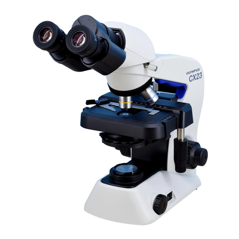
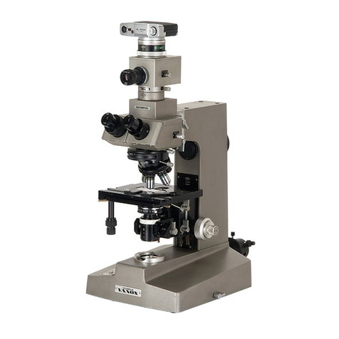
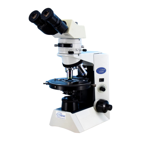
Fig.
1
-
--.
\q1
Explmatlons
In
detall
1.
MOUNTtMGTHE
HALOGEN
BULB
r~
1
Gently
insert
the
two
fineprongs
of
the
halogenbulb
into
thesmall holes
of
the lamp holder. Avoid fingerprints on the bulb.
Wipe
the bulb,
if
necessary.
2.
AllACHING
THE
LAMP
HOLDER
(Fig.
1)
-
FI~
the
lower
prongs
of
the
lamp
holder
and
the heavier
side
prongs
of
the lamp holder into the openings at the lower back
of
the
base
of
the
microscope.
3.
MOUNTING
THE
STAGE
{Fig.
2)
1)
Rack
down
the
stage
dwetail
El,
2)
Fit the
stage
carefully onto the dovetail. The Olympus nameplate
should
face
the
user.
The
two
stage
centering
screws
should
be
angledtoward
each
other;
one
to
theleft
of
center,theotherto
the
right
of
center,
3)
Tghtenthe stage
fastening
screw
@I.
This
screw
will be
found
on
the
user$
left
at
a
right
angle
to
the
body
of
the
microscope.
Rg.3
4.
MOUNTING
THE
CONDENSER
Loosen
the
condenser
clamping
screw.
Slidethe
condenser
in,
aperture
scale
facing
forward.
Tighten
the
clamping
screw.
5.
MOUNTING
THE
REVOLVING
NOSEPIECE
1)
Loosen
the
nosepiece
clamping
screw
Dl
(Rg.
3)
Fig.
4
2)
Aligning
the
nosepiece
dovetail
slide
El
to
themounting
block,
push
in
the
nosepiece
slowly
all
the
way.
Do
not
tilt
or
lock
the
nosepiecewhile inserting
into
themounting
block.
6.
MOUNTING
THE
INTERMEDIATEPOLARIZING
ATTACHMENT
(Fig.
4)
1)
Loosen
the
clamping
screw
El
fully.
Pull
spring-loadedclamping
screw
m.
This
will
cause
thelocatingpin
El
towithdraw.
(Fig.
4)
Ifthe
pin
does
not,
loosen
the
s~rew
further
until
the
pinwithdraws.
2)
Wfih
;lamping
screw
El
pulled
out,
insert
the
circular
dovetail
of
the
inlsrmediateattachment
into
the
ring
dwetait.
3)
Tghten
the
clamping
screw.
