Leica DM1000 LED User manual

Leica DM1000
Leica DM1000 LED
Operating Manual · Bedienungsanleitung · Mode d’emploi

Phone/Tel./Tél. +49 (0) 64 41-29 4253
Fax +49 (0) 64 41-29 2255
E-Mail/e-mail/courriel MQM-Hotline@leica-
microsystems.com
Published June 2010 by/
Herausgegeben Juni 2010 von/
Publiée en june 2010 par :
Leica Microsystems CMS GmbH
Ernst-Leitz-Strasse 17-37
D-35578 Wetzlar (Germany)
Responsible for contents/
Verantwortlich für den Inhalt/
Responsable du contenu rédactionnel :
Peter Schmitt, Manuela Jacobsen
(Clinical Imaging, Product Management)
Holger Grasse
(Safety Officer according to MPG §30/
Sicherheitsbeauftragter nach MPG §30/
Responsable de la sécurité conformément à la loi
relative aux dispositifs médicaux, §30)
In case of questions, please contact the hotline/
Bei Fragen wenden Sie sich bitte an die Hotline/
Pour toute question, contacter notre service
d’assistance téléphonique :

Leica DM1000
Leica DM1000 LED
Operating Manual

4
Copyrights
Copyrights
All rights to this documentation are held by
Leica Microsystems CMS GmbH. Reproduction
of text or illustrations (in whole or in part) by
print, photocopy, microfilm or other method (in-
cluding electronic systems) is not allowed with-
out express written permission from Leica
Microsystems CMS GmbH.
The instructions contained in the following
documentation reflect state-of-the-art technology.
We have compiled the texts and illustrations as
accurately as possible. Nevertheless, no liability
of any kind may be assumed for the accuracy of
this manual’s contents. Still, we are always
grateful for comments and suggestions
regarding potential mistakes within this
documentation.
The information in this manual is subject to modifi-
cation at any time and without notification.

5
Contents
8.3 Focusing ...................................................... 36
8.4 Tubes ....................................................... 37
8.5 Eyepieces.................................................... 38
8.6 Objectives ................................................... 39
8.7 Light Sources ............................................. 40
8.8 Aperture Diaphragm ................................. 40
8.9 Field Diaphragm ......................................... 41
9. Contrast Methods ...................................... 42
9.1 Transmitted Light ....................................... 42
9.1.1 Brightfield......................................... 43
9.1.2 Phase Contrast................................ 44
9.1.3 Darkfield ........................................... 44
9.1.4 Oblique Illumination ....................... 45
9.1.5 Polarization ...................................... 45
9.2 Fluorescence.............................................. 46
10. Measurements with the Microscope ... 47
10.1 Linear Measurements .............................. 47
10.2 Thickness Measurements ....................... 48
10.3 Differentiation of Gout / Pseudo Gout ... 49
11. Trouble Shooting ....................................... 51
12. Care of the Microscope ........................... 54
12.1 Dust Cover .................................................. 54
12.2 Cleaning....................................................... 54
12.3 Handling Acids and Bases ...................... 55
12.4 Changing Fuses.......................................... 55
13. Essential Wear and Spare Parts ............ 56
14. Retrofitting Components .......................... 57
14.1 Equipping the Condenser Disk ................ 57
15. Index ............................................................ 59
16. EC Declaration of Conformity ................. 60
Contents
1. Important Notes about this Manual ...... 6
2. Intended Purpose of the Microscope ... 7
3. Safety Notes ............................................... 8
3.1 General Safety Notes ............................... 8
3.2 Electrical Safety ........................................ 8
3.3 Disposal ....................................................... 9
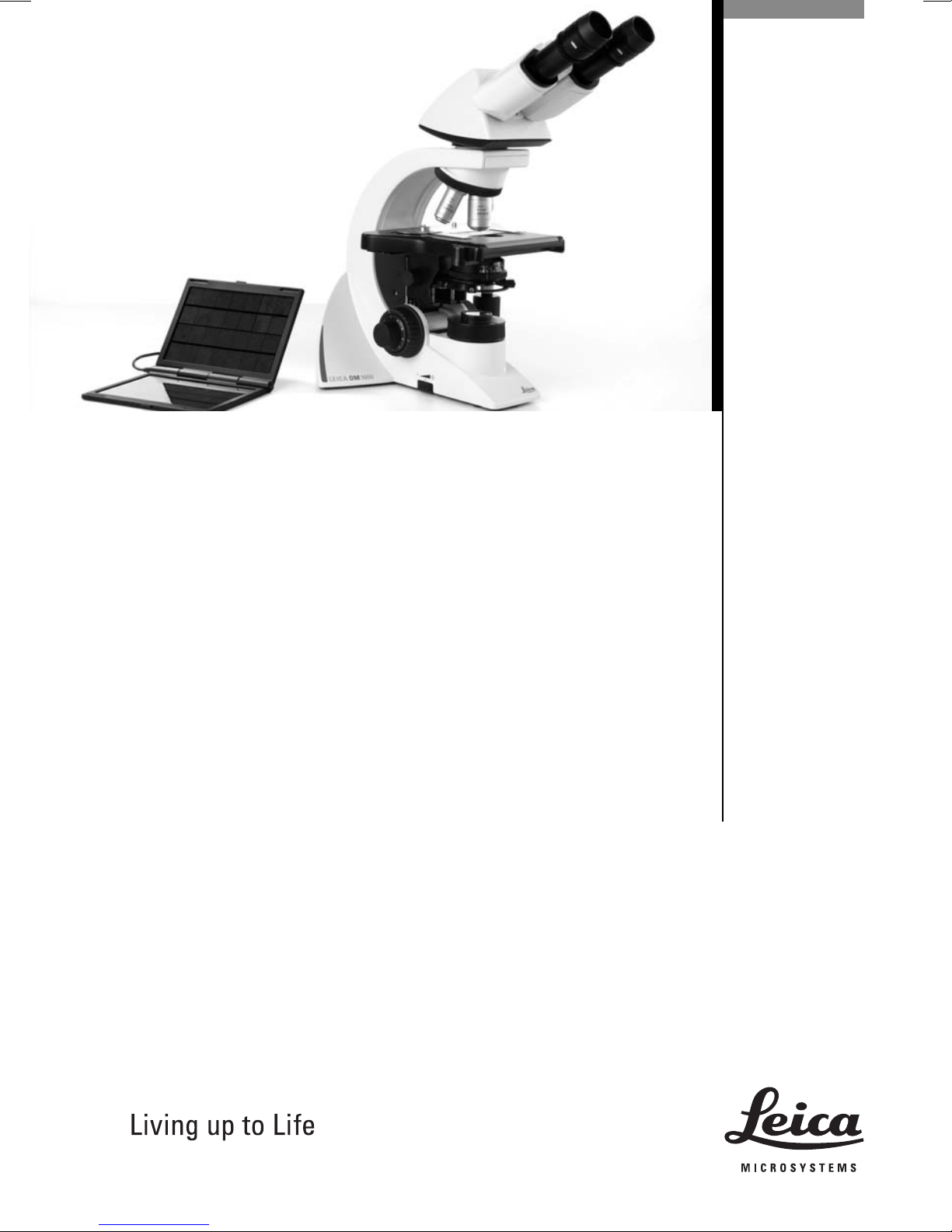
4. Overview of the Instrument .................... 10
5. Unpacking the Microscope .................... 14
6. Assembling the Microscope .................. 16
6.1 Stage ............................................................ 16
6.2 Condenser ................................................... 18
6.3 Tube and Eyepieces .................................. 19
6.4 Objectives ................................................... 19
6.5 Light Source - Transmitted Light Axis ... 20
6.6 Components for
Fluorescence Applications...................... 21
6.6.1 Fluorescence Illuminator .............. 21
6.6.2 106z Lamp Housing ......................... 21
6.7 Analyzer and Polarizer* .......................... 24
6.8 Lambda Plate Compensator .................... 24
6.9 Optional Accessories ............................... 25
6.10 Insertion of the batteries ......................... 27
6.11 Connection to the Power Supply............ 27
7. Startup ......................................................... 28
7.1 Switching on the Microscope................. 28
7.2 Köhler Illumination .................................... 28
7.3 Checking Phase Contrast Rings ............. 29
7.4 Adjusting the Light Sources .................... 31
8. Operation .................................................... 35
8.1 Switching on............................................... 35
8.2 Stages and Object Displacement........... 35

6
1. Important Notes about this Manual
(1.2)
→
p. 20
!
*
Numbers in parentheses, such as "(1.2)", corre-
spond to illustrations (in the example, figure 1,
item 2).
Numbers with pointer arrows (for example
→p. 20), point to a certain page of this manual.
Caution!
Special safety instructions within this
manual are indicated with the triangle
symbol shown here, and have a gray
background.
Caution! The microscope and accessories can
be damaged when operated incorrectly.
Notes on the disposal of the device,
accessories and consumable materials.
Explanatory note
Item not contained in all configurations
Text symbols, pictograms and their meanings:
Caution!
This operating manual is an essential com-
ponent of the microscope, and must be read
carefully before the microscope is
assembled, put into operation or used.
1. Important Notes about this Manual
This operating manual contains important in-
structions and information for the operational
safety and maintenance of the microscope and
accessories. Therefore, it must be kept and
taken care of.

7
2. Intended Purpose of the Microscope
The Leica DM1000/DM1000 LED microscope, to
which this user manual belongs, is designed for
biological routine and research applications.
This includes the examination of samples taken
from the human body with a view to provide
information on physiological or pathological
states or congenital abnormalities, or to
determine the safety and compatibility with po-
tential recipients, or to monitor therapeutic
measures.
The above-named microscope complies with
the Council Directive 98/79/EEC concerning in
vitro diagnostics. (It also conforms to the
Council Directives 2006/95/EC concerning
electrical apparatus and 2004/108/EC
concerning electromagnetic compatibility for
use in an industrial environment.)
Caution!
The manufacturer assumes no liability for
damage caused by, or any risks arising from
using the microscope for other purposes
than those for which they are intended or
not using them within the specifications of
Leica Microsystems CMS GmbH.
In such cases the conformity declaration
shall cease to be valid.
Caution!
These (IVD) devices are not intended for use
in the patient environment defined by DIN
VDE 0100-710. Neither are they intended for
combining with medical devices according
to EN 60601-1. If a microscope is electrically
connected to a medical device according to
EN 60601-1, the requirements defined in
EN 60601-1-1 shall apply.
Not suitable for examining potentially
infectious specimens.

8
3. Safety Notes
3. Safety Notes
3.2 Electrical Safety
General Specifications
Microscope
For indoor use only.
Supply voltage:
Power input:
Fuses:
Ambient temperature:
Relative humidity:
Over voltage category:
Pollution degree:
Specifications of the external power supply
ELPAC POWER SYSTEMS power supply,
Model: FW1812
Input: 100-240 V AC
0,5 A
47-63 Hz
Output: 12 V DC
1,5 A max.
18 W max.
Caution!
Use the original power supply only. Other
power supplies must not be used.
90-250 V AC, 50-60 Hz
(DM1000)
12 V DC, 1.5 A
(DM1000 LED)
40 W (DM1000)
18 W (DM1000 LED)
F 3.15 A 250 V
(DM1000)
Integrated in external
power supply unit,
not replaceable
(DM1000 LED)
15-35°C
max. 80% to 30°C
II
2
3.1 General Safety Notes
This safety class 1 (DM1000) or class 2
(DM1000 LED) device is constructed and tested
in accordance with
EN 61010-2-101:2002 (DM1000/DM1000 LED),
EN 61010-1:2001 (DM1000/DM1000 LED),
IEC 61010-1:2001 (DM1000/DM1000 LED),
IEC 60825-1:2007 (DM1000 LED)
EN 60825-1 + A1 + A2:2003 (DM1000 LED),
LED Class I (DM1000 LED)
safety regulations for electrical measuring, con-
trol, and laboratory devices.
Caution!
In order to maintain this condition and to en-
sure safe operation, the user must follow the
instructions and warnings contained in this
operating manual.
Caution!
The devices and accessories described in
this operating manual have been tested for
safety and potential hazards.
The responsible Leica affiliate or the main
plant in Wetzlar, Germany must be consulted
whenever the device is altered, modified or
used in conjunction with non-Leica compo-
nents that are outside of the scope of this
manual.
Unauthorized alterations to the device or
noncompliant use shall void all rights to any
warranty claims and product liability!

9
3. Safety Notes
If the original power supply fails or is
damaged, it must be replaced. Repair is not
permitted.
Original power supplies are available from
your Leica branch office or Leica dealer.
Caution!
The power plug may only be plugged into an
outlet equipped with a grounding contact.
Do not interfere with the grounding function
by using an extension cord without a ground
wire. Any interruption of the ground wire in-
side or outside of the device, or release of
the ground wire connection, can cause the
device to become hazardous. Intentional
ground interruption is not permitted!
Caution!
DM1000 only: Never use any fuses as
replacements other than those of the types
and the current ratings listed here. Using
patched fuses or bridging the fuse holder is
not permitted. The use of incorrect fuses
may result in a fire hazard.
Caution!
The microscope's electrical accessory com-
ponents are not protected against water.
Water can cause electric shock.
Caution!
Protect the microscope from excessive tem-
perature fluctuations. Such fluctuations can
lead to the accumulation of condensation,
which can damage the electrical and optical
components.
Ambient temperature: 15-35°C.
Caution!
DM1000 only: Before exchanging the fuses
or lamps, be absolutely certain to switch off
the main power switch and remove the po-
wer cable.
3.3 Disposal
To dispose of the product at the end of its ser-
vice life, please contact Leica Service or Sales.
Please observe national laws and regulations,
such as those implementing and enforcing the
WEEE EU directive.
Note:
Like other electronic devices, the micro-
scope and its accessory components must
not be disposed of as regular household
waste.

10
4. Overview of the Instrument
Specification
Contrast Methods
Transmitted Light Axis
Incident Light Axis
(optional)
Tube
Magnification Changer
(optional)
Objective Turret
X/Y Stage
Leica DM1000/DM1000 LED
• Transmitted light: brightfield, darkfield,
phase contrast, polarization
• Incident light: fluorescence
Leica DM1000: Integrated halogen illumination
Leica DM1000 LED: Integrated LED illumination
manual adjustment of
• Light intensity
• Aperture diaphragm
• Field diaphragm (only with Köhler kit)
Incident light fluorescence illuminator for eyepieces with
field number up to 20 with
• Interchangeable slide with mount for 3 filter systems
• Adjusting lens for lamp
• Light trap for the suppression of extraneous light
• BG38 blue filter and shutter, switchable
optionally with
• Fixed or variable viewing angle
• Up to 3 switching positions
• one or two camera ports
• Ergotube with height-adjustable eye level and camera port
• Manual
• Magnification steps: 1x; 1.5x; 2x
• Manual
• 5-fold for objectives with M25 thread
• With condenser holder
• Coaxial pinion, optional telescopable
• Controls mountable left or right

11
4. Overview of the Instrument
Specification
Condenser
Focusing
Leica DM1000/DM1000 LED
optionally with
• CL/PH 0.90/1.25 OIL condenser with color coding
• CLP/PH 0.85 condenser for polarization
• Achr.apl. A 0.9 (P) condenser with swivelable condenser head
• UCL 0.90/1.25 OIL universal condenser UCLP 0.85 for
polarization with 5-position light ring disk)
• UCL/P pol. universal condenser with interchangeable
condenser head and condenser disk with 6 positions
• Focus wheel for coarse and fine focusing
• Height adjustment

12
4. Overview of the Instrument
Fig. 1 Left side of the Leica DM1000 stand
1Coarse and fine focusing
2Condenser height adjustment
3Brightness control
4Field diaphragm
5Aperture diaphragm
6Condenser
6
5
4
12 3

13
4. Overview of the Instrument
Fig. 2 Right side of the Leica DM1000 stand
1Eyepiece
2Eyepiece tube
3Tube
4Objective turret with objectives
5Specimen stage with specimen holder
6 Integrated illumination
7On/Off switch
8Condenser height adjustment
9Coarse and fine focusing
10 Coaxial pinion for x/y stage movement
1
2
3
4
5
678910

14
5. Unpacking the Microscope
Installation Location
Work with the microscope should be performed
in a dust-free room, which is free of vapors (oil,
chemicals etc.) and of extreme humidity. At the
workplace, large temperature fluctuations,
direct sunlight and vibrations should be avoided.
These conditions can distort measurements and
micrographic images.
Allowable ambient conditions
Temperature 15-35°C
Relative humidity maximum 80% up to 30°C
Microscopes in warm and warm-damp climatic
zones require special care in order to prevent
the build up of fungus.
See the chapter "Care of the Microscope" →
p. 54,
for additional instructions.
Caution!
Electrical components must be placed at least
10 cm away from the wall and away from
flammable substances.
First, carefully remove all components from the
transportation and packaging materials.
Note:
If at all possible, avoid touching the lens sur-
faces of the objectives. If fingerprints do appear
on the glass surfaces, remove them with a soft
leather or linen cloth. Even small traces of finger
perspiration can damage the surfaces in a short
time. See the chapter "Care of the Microscope"
→
p. 54, for additional instructions.
Caution!
Do not yet connect the microscope and pe-
ripherals to the power supply at this point!
5. Unpacking the Microscope

15
5. Unpacking the Microscope
Transport
For shipping or transporting the microscope
and its accessory components, the original
packaging should be used.
As a precaution to prevent damage from vibra-
tions, the following components should be dis-
assembled and packaged separately:
• Unscrew the objectives
• Remove the condenser
• Remove the coaxial pinion
• Remove the lamp housings
• Disassemble the burner of 106z lamp housing
• Remove all moving or loose parts

16
6. Assembly
!Fig. 3 Specimen stage with specimen holder
1Lock screws for specimen holder
The microscope components are logically as-
sembled in this order:
• Stage
• Condenser
• Fluorescence illuminator*
• Intermediate systems*
• Tube
• Eyepieces
• Objectives
• Lamp housings with light sources*
• Polarization equipment*
Only one commonly used screwdriver is
necessary for assembly, which is included in the
delivery package.
The tool can be stored on a magnetic retainer
on the underside of the stage at the right.
When using intermediate systems and optical
accessories, the sequence may vary.
In this case, read Chapter
"6.9 Optional Accessories" →p. 25.
6.1 Stage
Caution:
Before completing the stage, make sure no ob-
jectives are installed!
Remove the srew located under the stage in the
front.
Specimen Holder
• Place the specimen holder on the stage and
fasten it with the two screws (3.1)
Coaxial Pinion
Note:
The coaxial pinion can be mounted on the left-
or right-hand side.
• First, place the flat fine focus wheel on the
side to which you intend to mount the coaxial
pinion; the wheel is held in place magnetically
(4.1); ensure that the button snaps into place.
Attach the other focus knob on the opposite side
• Loosen the lock screw (5.1) at the front left-
hand side of the stage
• Slide the stage as far back as possible
• Attach the coaxial pinion with the srew (6.1)
• Return the stage to the starting position and
retighten the lock screw; after installation of
the stage control, move object guide all the
way to the left side of the instrument; keep
turning when guide has reached the end of
travel until a click noise is heard
1
6. Assembling the Microscope

17
6. Assembly
Adjusting the Focus Stop
The focus stop is preadjusted by the factory to
prevent collision with objectives. The focus stop
should be set approx. 0.3 mm higher as the focal
plane to enable focussing of the samples of dif-
ferent thickness.
Fig. 6 Coaxial pinion installation
1Mounting screw for coaxial pinion
1
1
Fig. 4 Focus wheel
1Magnetic retainer for fine focus wheel
Fig. 5 Underside of stage
1Lock screw
1
If a readjustment is necessary, adjust as
following:
• Lower the stage by rotating 1/2 turn of the
coarse adjustment knob
• Loosen the focus stop screw on left hand side
of microscope
• Move stage to desired focal plane (preferably
approx. 0.3 mm higher)
• Tighten focus stop screw
Stage Lock*
The stage lock is mounted in the same hole as
the stage drive (in case of ErgoStages, it can be
mounted on the opposite position in addition to
stage drive).
The mounting of stage lock is similar to
mounting of the stage drive:
• Loosen the locking screw (5.1) on the left side
underside of stage and move the stage all the
way back
• Loosen the screw for coaxial pinion (6.1),
remove the coaxial pinion, and attach the
stage lock with this screw
• Loosen the stage lock screw and pull the
stage forward to the desired position
• Retighten the locking screw (5.1) underside of
stage
• Press the pinion of the stage lock against the
rack and retighten the stage lock screw

18
6. Assembly
6.2 Condenser
• If present, screw the condenser head into the
condenser
• Using the condenser height adjuster (9.3), turn
the condenser holder (fig. 8) completely
downward
• Unscrew the clamping screw for the con-
denser (9.2) far enough so that the condenser
can be inserted from the front
• From the front, insert the condenser into the
condenser holder as far as it will go; on the
underside of the condenser, there is an orien-
tation pin (7.1), which must be located in the
guiding notch (8.1)
• Pull the condenser's clamping screw (9.2) so
that the condenser is locked in place
Fig. 9 Condenser holder
1Condenser centering bolts
2Clamping screw for condenser
3Condenser height adjuster
Fig. 8 Condenser holder
1Guiding notch
1
Note:
The condenser must be centered before using
the microscope.
→
Köhler illumination p. 28.
Fig. 7 Underside of condenser (example CL/PH)
1Orientation pin
2Auxiliary condenser lens LS
2
11
3
2
1
3
1

19
6. Assembly
6.3 Tube and Eyepieces
Note:
For fluorescence applications, install the
fluorescence illuminator* first →p. 21.
If available, the analyzer* (10.1) must be
inserted into the stand. This requires that the
guide key engages in the guide pin (10.2).
To mount the analyzer, the analyzer mount TL*
20 mm or 60 mm can also be placed between
stand and tube.
An intermediate tube pole* with a switchable
analyzer (on/off) and Bertrand lens is also
available as an option.
The tube is mounted to the stand either directly or
with the use of intermediate modules*.
Fig. 11 Fastening the tube
1Clamping screw
1
!
Fig. 10 Analyzer mounting
1Analyzer
2Orientation pin and guiding notch
3Clamping screw
• Loosen the clamping screw (11.1) on the
stand
• Insert the tube in the circular receptacle
(dovetail ring)
• Retighten the clamping screw (11.1)
• The eyepieces are inserted into the eyepiece
tubes on the tube
6.4 Objectives
Always only use Leica objectives of tube length ¥
(infinity)! The standard thread is M25. The
objectives should be arranged so that the
magnification increases when the objective
nosepiece is rotated counter-clockwise.
Attention:
Lower the specimen stage as far as possible
before assembling the objectives. Close vacant
threads in the nosepiece with dust protection
caps!

20
6. Assembly
6.5 Light Source for the Transmitted Light Axis
Note:
The Leica DM1000 LED is equipped with
integrated LED illumination. The service life of
the LED is about 100.000 hours. If, despite this, it
should be necessary to change the LED, this
task must be carried out by Technical Service
only.
The following instructions in this chapter refer to
the Leica DM1000 with tungsten halogen lamp.
Caution!
Be sure that the microscope and lamp
housing are disconnected from the power
supply. Unplug the power plug and the po-
wer supply during assembly.
Caution!
Light sources pose a potential irradiation
risk (glare, UV-radiation, IR-radiation).
Therefore, lamps have to be operated in
closed housings.
Replacing the Lamp of the Integrated
Illumination
The transmitted light illumination with a low-
voltage tungsten halogen lamp (fig. 12) is
integrated in the base of the microscope and is
accessible from the right-hand side.
• Remove the insert (12.2)
Caution!
The lamp may still be hot!
• Remove the lamp
Caution!
Do not remove the new lamp's dust cover
until you have installed the lamp. Avoid
fingerprints on the lamp.
• Insert the new lamp with the dust cover
straight into the socket until it stops; be sure
that the lamp is inserted straight
• Remove the lamp's dust cover
• Replace the insert (12.2)
Fig. 12 Transmitted-light illumination
in Leica DM1000 microscope base
1Tungsten halogen lamp
2Insert
12
Other manuals for DM1000 LED
2
Table of contents
Languages:
Other Leica Microscope manuals

Leica
Leica DM750 M User manual

Leica
Leica DMI Series User manual

Leica
Leica ATC 2000 User manual

Leica
Leica TCS SP2 Reference guide

Leica
Leica DM IRB User manual

Leica
Leica FL800 ULT User manual

Leica
Leica DMS1000 User manual

Leica
Leica SP2 User manual

Leica
Leica ATC 2000 User manual

Leica
Leica S Series User manual